Hookworm

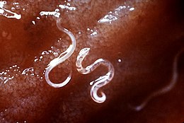
Ancylostoma caninum hookworms in a dog
Hookworms are intestinal, blood-feeding, parasitic roundworms that cause types of infection known as helminthiases. In humans, hookworm infections are caused by two main species of roundworm belonging to the genera Ancylostoma, and Necator. In other animals the main parasites are species of Ancylostoma.
Contents
1 Species
2 Characteristics
2.1 Life cycle
3 References
Species

Infective N. americanus larva
The two most common types of hookworm that infect humans are Ancylostoma duodenale, and Necator americanus.
Hookworm species that are known to infect cats are Ancylostoma braziliense, and Ancylostoma tubaeforme. Wild cats are infected by Ancylostoma pluridentatum.
Dogs are commonly infected by Ancylostoma caninum.
The only zoonotic hookworm is Ancylostoma ceylanicum that can infect humans and other mammals.[1]
Characteristics
The two common species that infect humans share a similar morphology. A. duodenale worms are pale grey or slightly pink. The head is bent a little in relation to the rest of the body, forming a hook shape – hence the name. The hook is at the front end of the body. They have well-developed mouths with two pairs of teeth. Males measure approximately one centimeter by 0.5 millimeter, and females are often longer and stouter. Males also have a prominent copulatory bursa posteriorly.[2]
N. americanus is generally smaller than A. duodenale with males usually 5 to 9 mm long and females about 1 cm long. Instead of the two pairs of teeth in A. duodenale, N. americanus has a pair of cutting plates in the buccal capsule. Also the hook is much more defined in Necator americanus.[2]
Life cycle

Hookworm life cycle
The host is infected by the larvae, not by the eggs and the usual route is through the skin. Hookworm larvae need warm, moist soil, above 18°C in order to hatch. They will die if exposed to direct sunlight or if they become dried out. Necator larvae can survive at higher temperatures than Ancylostoma larvae.
First stage larvae (L1) are non-infective, and once hatched into the deposited feces feed on this, and then feed on soil microorganisms until they moult into second stage larvae (L2).[3] First and second stage larvae are in the rhabditiform stage. After feeding for around seven days or so they will moult into third stage larvae (L3) known as the filariform stage. This is the non-feeding, infective stage. Filariform larvae can survive for up to two weeks, they are extremely motile and will move onto higher ground to improve their chances of finding a host. N. americanus larvae can only infect through penetrating skin, but A. duodenale can also infect orally. A common route of passage for the lavae is the skin of barefoot walkers. Once the larvae have entered the host they travel in the circulatory system to the lungs where they leave the venules and enter the alveoli. They then travel up the trachea where they are coughed up, swallowed and end up in the small intestine. In the small intestine the larvae moult into stage four (L4) the adult worm. It takes from five to nine weeks from penetration to maturity in the intestine.[4][5]
Necator americanus can cause a prolonged infection lasting from one to five years with many worms dying in the first year or two. Some worms though have been recorded as living for fifteen years or more. In comparison, Ancyclostoma duodenale worms are short-lived lasting for around six months. However, larvae can remain dormant in tissue stores and be recruited over many years to replace the worms that die.
The worms mate inside the host, and the females lay their eggs, to be passed out in the host’s feces into the environment to start the cycle again. N. americanus can lay between nine and ten thousand eggs per day, and A. duodenale between twenty-five and thirty thousand per day. Their eggs are indistinguishable.
Worms need five to seven weeks to reach maturity and symptoms of infection can therefore appear before eggs are to be found in the feces, making a diagnosis of hookworm infection difficult.
References
^ Traub, RJ (November 2013). "Ancylostoma ceylanicum, a re-emerging but neglected parasitic zoonosis". International Journal for Parasitology. 43 (12–13): 1009–15. doi:10.1016/j.ijpara.2013.07.006. PMID 23968813..mw-parser-output cite.citation{font-style:inherit}.mw-parser-output .citation q{quotes:"""""""'""'"}.mw-parser-output .citation .cs1-lock-free a{background:url("//upload.wikimedia.org/wikipedia/commons/thumb/6/65/Lock-green.svg/9px-Lock-green.svg.png")no-repeat;background-position:right .1em center}.mw-parser-output .citation .cs1-lock-limited a,.mw-parser-output .citation .cs1-lock-registration a{background:url("//upload.wikimedia.org/wikipedia/commons/thumb/d/d6/Lock-gray-alt-2.svg/9px-Lock-gray-alt-2.svg.png")no-repeat;background-position:right .1em center}.mw-parser-output .citation .cs1-lock-subscription a{background:url("//upload.wikimedia.org/wikipedia/commons/thumb/a/aa/Lock-red-alt-2.svg/9px-Lock-red-alt-2.svg.png")no-repeat;background-position:right .1em center}.mw-parser-output .cs1-subscription,.mw-parser-output .cs1-registration{color:#555}.mw-parser-output .cs1-subscription span,.mw-parser-output .cs1-registration span{border-bottom:1px dotted;cursor:help}.mw-parser-output .cs1-ws-icon a{background:url("//upload.wikimedia.org/wikipedia/commons/thumb/4/4c/Wikisource-logo.svg/12px-Wikisource-logo.svg.png")no-repeat;background-position:right .1em center}.mw-parser-output code.cs1-code{color:inherit;background:inherit;border:inherit;padding:inherit}.mw-parser-output .cs1-hidden-error{display:none;font-size:100%}.mw-parser-output .cs1-visible-error{font-size:100%}.mw-parser-output .cs1-maint{display:none;color:#33aa33;margin-left:0.3em}.mw-parser-output .cs1-subscription,.mw-parser-output .cs1-registration,.mw-parser-output .cs1-format{font-size:95%}.mw-parser-output .cs1-kern-left,.mw-parser-output .cs1-kern-wl-left{padding-left:0.2em}.mw-parser-output .cs1-kern-right,.mw-parser-output .cs1-kern-wl-right{padding-right:0.2em}
^ ab Markell, Edward K.; John, David C.; Petri, William H. (2006). Markell and Voge's medical parasitology (9th ed.). St. Louis, Mo: Elsevier Saunders. ISBN 978-0-7216-4793-7.
^ "Ancylostoma duodenale". Animal Diversity Web.
^ Hotez, PJ; Bethony, J; Bottazzi, ME; Brooker, S; Buss, P (March 2005). "Hookworm: "the great infection of mankind"". PLoS Medicine. 2 (3): e67. doi:10.1371/journal.pmed.0020067. PMC 1069663. PMID 15783256.
^ "CDC Factsheet: Hookworm" Archived 2010-09-04 at the Wayback Machine